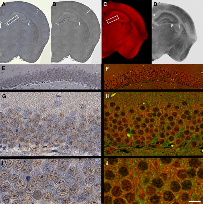
Figure 3.

IR fluorophores are compatible with higher resolution imaging by confocal microscopy. Representative tissue sections immunostained for D2 receptor probed with conventional DAB labelling (A, B, E, G, I) or IRDye 680 (C, D, F, H, J). (A, B) Whole coronal reconstructed tissue sections are shown in colour and grey‐scale. (C, D) IR‐captured tissue sections in colour and grey‐scale, respectively. (A, C) An area of the DG outlined with a white rectangular box, subsequently imaged at higher magnification (20 × e and f; 63 × g and h) on either a light or confocal microscope (e and g; f and h, respectively). (I and J) Close crops taken from (G and H) using adobe photoshop. Scale bar: 1 mm (A–D); 80 μm (E and F); 20 μm (G and H); 10 μm (I and J).
