Figure 4.

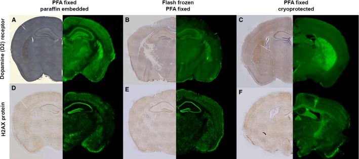
Protein distribution assessed by IR QFIHC is comparable to DAB reporter staining regardless of upstream tissue processing methodologies. The effect of fixation on tissue sections immunostained for two well‐characterised proteins, the dopamine (D2) receptor (A–C) and the nuclear protein H2A.X (D–F). Sections were imaged using either conventional DAB‐based IHC light microscopy or IR‐based scanning. Different tissue fixation methodologies are shown with: (A, D) tissues fixed in paraformaldehyde (PFA) followed by paraffin‐embedding; (B, E) tissues were flash frozen prior to sectioning followed by paraformaldehyde fixation; and (C, F) tissues have been fixed with paraformaldehyde prior to being stored in a cryopreservative and then sectioned. Each panel consists of DAB‐labelled hemispheres (LHS) and IR Dye 800 probed hemispheres (RHS). Scale bar: 1 mm.
