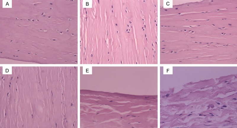
Figure 1.

Histological characteristics of lateral meniscus by H&E staining. A. In the control side, collagen fibers and meniscal cell morphology were normal. Meniscal cells were oval or fusiform with big and round nucleus. Collagen fibers were thick and orderly aligned. B. On 4 weeks after PCL-rupture, the H&E staining showed integrate surface structure and thick, loose and orderly arranged collagen fibers. C. On 8 weeks after PCL-rupture, the H&E staining showed integrate surface structure and thick, loose and orderly arranged collagen fibers. D. On 12 weeks after PCL-rupture, H&E staining showed rough surface structure with dent, and loose, orderly aligned and unevenly thick collagen fibers. Meniscal cells appeared disorderly arrangement and decreased in number. E. On 16 weeks after PCL-rupture, H&E staining appeared rough surface structure with dent, and unevenly thick, disorderly arranged collagen fibers. Meniscus cells appeared disorderly arrangement and obvious decreased in number. F. On 24 weeks after PCL-rupture, H&E staining showed fracture surface, and loose, unevenly thick and disorderly arranged collagen fibers. Cartilage cells were rarely visible.
