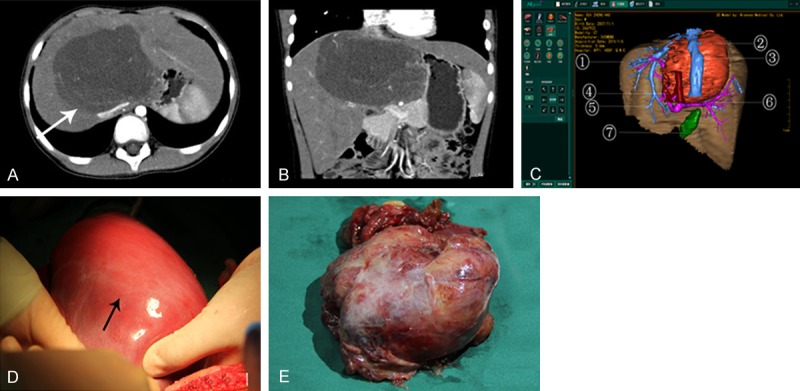
Figure 2.

Medical records of one case with tumor in the middle lobe of liver. A. The arrow in ordinary contrast-enhanced CT image indicates a tumor in the middle lobe; B. 3D reconstructed image using multiplanar reconstruction (MPR) shows the rough position of tumor. However, the precise anatomical relationship between tumor, portal vein and its branches, hepatic artery and hepatic vein could not be visualized. The success rate of surgery was predicted only approximately; C. 3D reconstructed image using Higemi clearly shows the boundaries of tumor and the neighboring relationship with blood vessels: ① liver, ② tumor, ③ inferior vena cava, ④ hepatic artery, ⑤ hepatic vein, ⑥ portal vein, ⑦ gall bladder; D. The arrow indicates the tumor in the middle lobe in intraoperative finding; E. Gross specimen of tumor, pathologically classified as malignant mesenchymoma with high malignancy and low differentiation.
