
Figure 2.
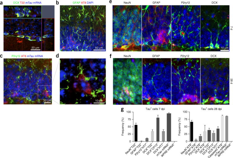
Immunofluorescence for tau with cellular markers. (a) Stacked sequential confocal microscopy imaging at 7 dpi of the GCL in the C57BL/6 mouse brain injected with AAV-GFP/tau into the MEC. DCX, green; T22, red; DAPI, blue. Scale bar, 20 μm. (b,c) Immunofluorescence at 28 dpi of the GCL in the C57BL/6 mice brain injected with AAV- GFP/tau in the MEC: GFAP (astrocytes, green), AT8 (red) and DAPI (blue) or P2ry12 (microglia, green), AT8 (red) and mTau mRNA (in situ hybridization, blue). (d) Stacked sequential confocal microscopy imaging of sections in c. AT8+ and mTau mRNA+ tau-bearing neurons are surrounded by P2ry12+ microglia (green). Scale bar, 10 μm. (e,f) Immunofluorescence of the GCL of a AAV-GFP/tau-injected mouse brain for cellular markers (green) and tau markers (red) at 7 (e) or 28 dpi (f). Scale bar, 10 μm. (e) From left to right, NeuN (mature neurons) and T22, GFAP and T22, P2ry12 and HT7 (hTau), and DCX and T22. (f) NeuN and AT8, GFAP and AT8, P2ry12 and AT8, and DCX and AT8. (g) Quantification of tau+ cells in AAV-GFP/tau-injected mice at 7 (left) or 28 dpi (right). The values represent double-positive cells with the cellular marker and the tau marker as a percentage of total tau marker–positive cells in the DG (mean ± s.e.m.). For mTau and WPRE mRNA, in situ hybridization was performed before the immunostaining for tau markers (T22 for 7 dpi and AT8 for 28 dpi).