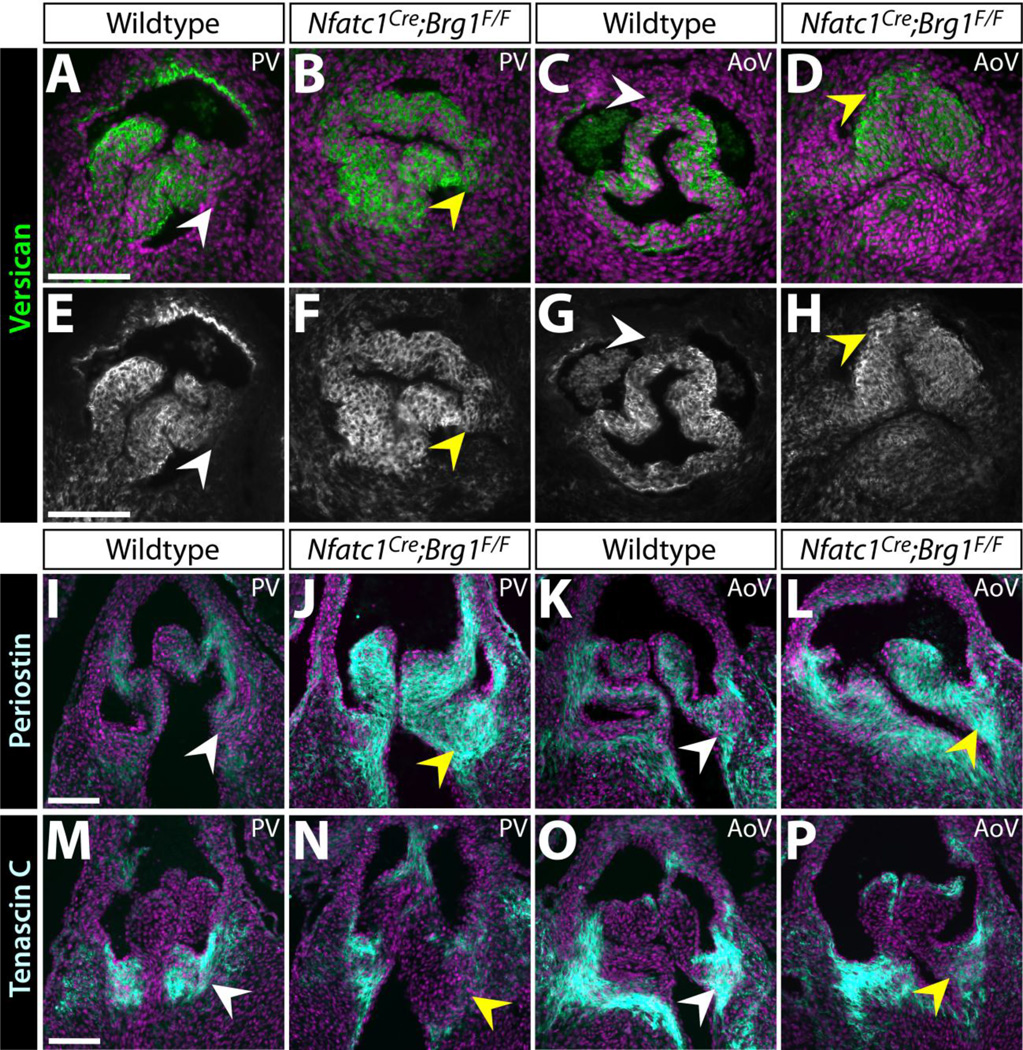
Figure 3. Altered localization and levels of extracellular matrix proteins in Nfatc1Cre;Brg1F/F semilunar valves.
(A–H) Widefield fluorescence images of anti-Versican (Vcan) antibody stained sections of E16.5 wildtype and Nfatc1Cre;Brg1F/F embryos. (I–P) Representative confocal imaged cryosections of E16.5 wildtype and Nfatc1Cre;Brg1F/F semilunar valves fluorescently immunostained for Periostin (Postn) (I–L) or Tenascin C (Tnc) (M–P) (n=3, paired control and Brg1-deficient valves). Pulmonic (PV) or aortic (AoV) valves are shown as indicated. ECM proteins are shown in green/gray scale (A–D) or cyan (I–P). Hoechst-stained nuclei are blue. White arrowheads mark areas of normal protein expression. Yellow arrowheads show regions with broadened (Vcan), broadened/increased (Postn) or decreased (Tnc) levels. Scale bars: 100 µm.

