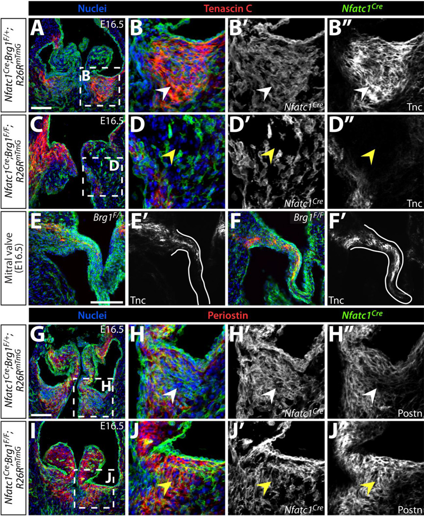
Figure 4. Disorganized extracellular matrix in endocardial Brg1 deficient semilunar valves reflects an altered organization of distinct mesenchymal lineages.
Immunofluorescence images of E16.5 control (Nfatc1Cre;Brg1F/+;R26RmTmG) or endocardial Brg1-deficient (Nfatc1Cre;Brg1F/F;R26RmTmG) embryo sections simultaneously stained for GFP to monitor Nfatc1Cre-lineage cells and either Tenascin C (Tnc) (A–F) or Periostin (Postn) (G–J). Pulmonic valves (A–D, G–J) and mitral valves (E–F) are shown. All panels are confocal images except (E–F), which are widefield images. Overlay images show Tnc or Postn in red, GFP indicating Nfatc1Cre-lineage cells in green, and Hoechst-stained nuclei in blue. Magnified regions are outlined in dashed boxes. White arrowheads denote normal ECM localization and yellow arrowheads represent either deficient (Tnc) or ectopic (Postn) ECM protein. Scale bars: 100 µm.

