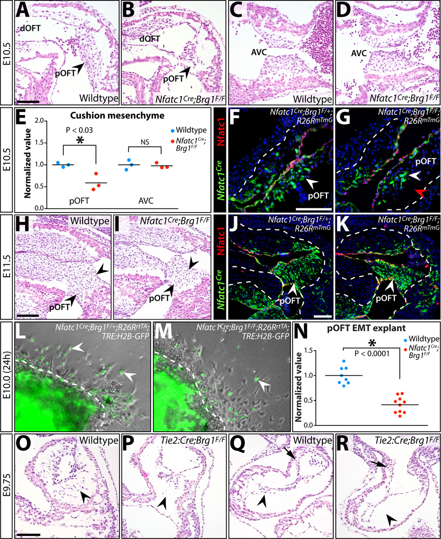
Figure 6. Reduced EMT-derived semilunar valve mesenchyme i7n Nfatc1Cre;Brg1F/F embryos originates from deficient pOFT EMT, a process that generally requires Brg1 function.

(A–D) H&E stained sagittal sections of E10.5 wildtype and Nfatc1Cre;Brg1F/F embryos. The proximal (pOFT) and distal (dOFT) outflow tract (A, B) and atrioventricular canal (AVC) cushions (C, D) are shown. Arrowheads indicate pOFT mesenchyme. (E) Scoring of mesenchymal cells populating the pOFT or AVC cushions in E10.5 wildtype and Nfatc1Cre;Brg1F/F embryos. Each point shows one embryo and all values are normalized to the mean of littermate wildtype samples. Asterisk denotes a significant difference (two-tailed Student’s t-tests). (F-G, J-K) Widefield fluorescence images of OFT regions from transverse sectioned E10.5 (F, G) or sagittal sectioned E11.5 (J, K) embryos. Tissue is immunostained for GFP (for Nfatc1Cre-lineage tracing, green) and Nfatc1 (endocardium, red). Nuclei are stained with Hoechst (blue). Control (Nfatc1Cre;Brg1F/+;R26RmTmG; F, J) and Nfatc1Cre;Brg1F/F;R26RmTmG (G, K) embryos are shown. (H, I) H&E stained sagittal sections of E11.5 wildtype and Nfatc1Cre;Brg1F/F embryos. Arrowheads indicate pOFT mesenchyme. (L, M) Brightfield and fluorescence overlaid images of collagen gel OFT explants from E10.0 control and Nfatc1Cre;Brg1F/F embryos (24 hours post-dissection). All embryos additionally carry R26RrtTA and TRE:H2B–GFP transgenes and explants are treated with doxycycline to induce H2B–GFP expression (to label Nfatc1Cre-lineage mesenchymal cells, green). White arrowheads denote EMT-derived (GFP+) mesenchymal cells. (N) Quantification of Nfatc1Cre-lineage mesenchymal cells migrating through the collagen matrix. Each point represents one explanted OFT. Values are normalized to the mean of littermate wildtype explants. The asterisk indicates statistical significance (two-tailed Student’s t-test). (O–R) Endocardial Brg1 is required for all cushion EMT. H&E stained sections showing AVC (O,P) and pOFT (Q,R) cushions of wildtype and Tie2:Cre;Brg1F/F E9.75 littermate embryos. Arrowheads denote either the presence or a depleted/absent pool of endocardial-derived cushion mesenchymal cells. Arrows in (Q, R) show neural crest-origin mesenchyme within the dOFT cushions. Scale bars: 100 µm.
