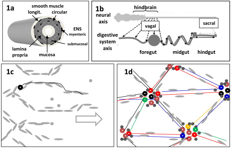
Fig. 1.
Diagrams of the structure and development of the ENS. (a) The ENS forms two 2-dimensional arrays (myenteric and submucosal plexuses) in the wall of the gut, associated with the intestinal smooth muscle. (b) Colonization of the foregut, midgut and hindgut in succession by vagal NC cells derived from the embryonic hindbrain. Sacral NC cells have a minor contribution to hindgut ENS. In Hirschsprung disease, the wave of vagal-derived ENC cells fails to complete the final occupation of the distal hindgut. (c) En face overview of the ENS at an early stage near the wavefront of colonization showing chain migration of ENC cells (grey) in the overall direction of the arrow. In mouse but not chick, some early-differentiating neurons (black circle) extend axons (black line) paralleling the ENC cell chains. (d) The same region after initial ganglionation, showing the geometric arrangement of the ENS ganglia. Neurons (colored circles representing different neuron types) form clusters and glial/ENC cells (grey) tend to surround the neurons. Nerve fibers (colored lines) accompanied by glial cells link the ganglia.

