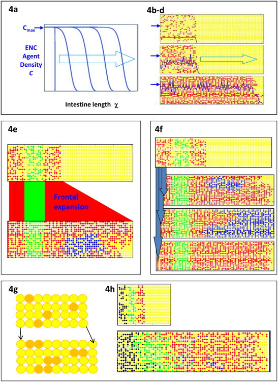
Fig. 4.
Simulations of the occupation of the gut by ENC agents. (a) A continuum model with diffusion-like movement and proliferation of ENC agents (dark blue trace) up to a set carrying capacity Cmax (dark blue arrow). This generates a directional wave (light blue arrow) of colonization. (b-d) CA model of ENC agent (red) occupation of the gut field (yellow). Dark blue line represents the density of ENC agents, and the dark blue arrows indicate the nominal carrying capacity Cmax. ENC agents are initially placed at density below Cmax Colonization of unoccupied gut is negligible until local ENC agent density rises by proliferation to approach Cmax. After this the ENC agents colonize the gut as a directional wave. Stochastic variation of movement and proliferation means that, in a CA simulation, the achieved density C varies continually around Cmax, unlike the agent density in the continuum model. (Images: M. Simpson). (e) Arbitrarily color coding regions of the ENC agent population shows that the front phalanx of ENC agents expands by proliferation to colonize the remaining unoccupied gut field (yellow). Two frontal ENC agents are labeled blue; their descendants form compact clones which in this particular simulation have merged. (Image: B. Binder) (f) In a partially colonized gut field (as for Fig. 4e), two frontal ENC agents are labeled blue (top panel); and migration and proliferation proceed under the basic conditions until the field is fully colonized. With identical stating conditions, stochastic variation in agent motility and proliferation results later in widely varying clonal outcomes. Some clones are moderate sizes (2nd panel) but some huge clones occur (3rd panel), as do clones with minimal or no expansion (bottom panel). This enormous range of clone sizes is an emergent property of this model that has not yet been investigated biologically. (Image: B. Binder). (g) The gut mesenchyme agent field (yellow) can be coded to grow in CA models. Individual mesenchyme agents in each row (g, top) are chosen at random to divide (orange) with daughter cell placed one site distal (to the right). In this way the gut field (g, bottom) elongates by one column per division cycle. (h) ENC agents on this growing field still show frontal expansion (red agents) as in non-growing fields (see Fig. 4e) but there is considerable population expansion behind the wavefront and mixing of ENC agents (green, black ENC agents). In addition, ENC clones (blue agents) do not remain compact when the field is growing. (Image: B. Binder)

