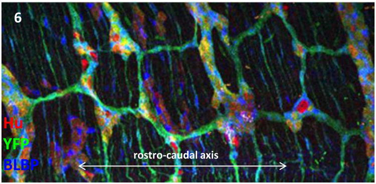
Fig. 6.
ENS cells re-organise to form a geometric array of ganglia and connecting tracts. YFP (green) reveals genetically marked ENS cells in wholemount of the mouse midgut at postnatal day 20. Neurons (Hu, red) and glial cells (BLBP, blue) cells associate in a pattern that is set early in development. (Image: Jun Lei)

