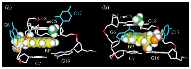
Figure 8.
Base-displaced intercalative conformations. (a) Representative NMR structure of the 10R (−)-trans-anti-[BP]G adduct, and (b) a modeled 10S (+)-trans-anti-[BP]G adduct (see the text), all in the (meC5-[BP]G6-C7)·(G16-C17-G18) sequence context. The selectively displayed protons are highlighted as white spheres, the carbon atoms of benzo[a]pyrene (BP) ring are highlighted as yellow spheres, the modified guanine residue and its opposing cytosine residue are shown as stick models (cyan), the carbon atom of the methyl group of the meC5 residue is shown as a green sphere, the oxygen atoms of the benzylic hydroxyl groups are shown as orange spheres.

