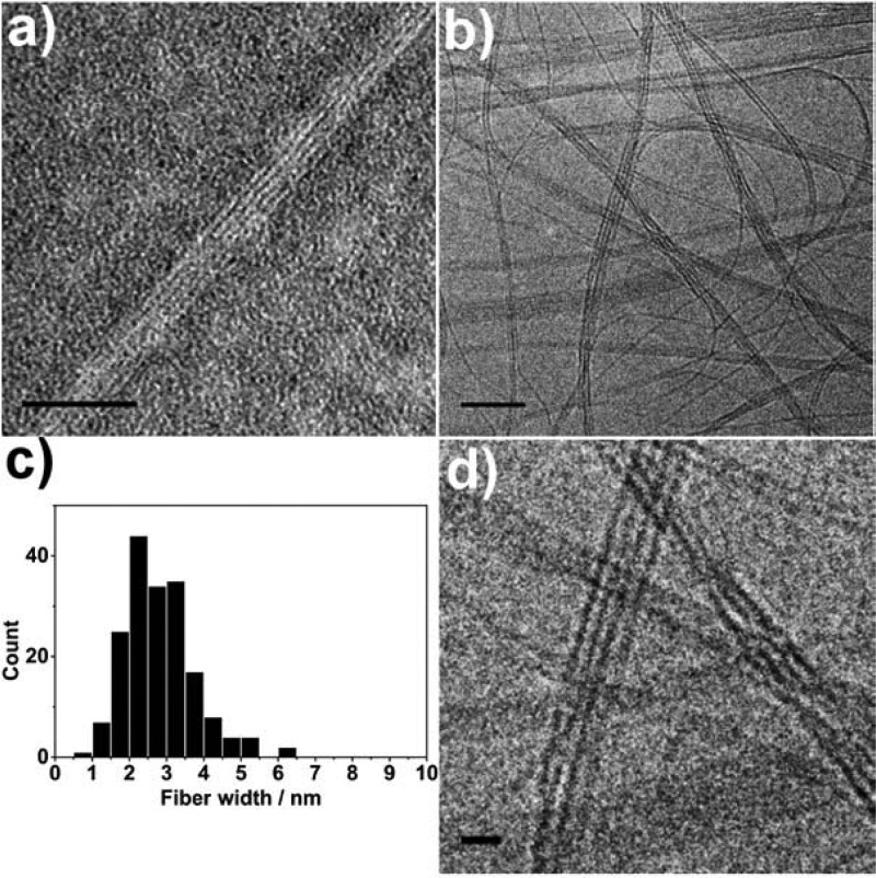
Figure 2.
(a) TEM (stained with 1% uranyl acetate) and (b) cryo-TEM (unstained) of EB TANI-PTAB forming bundles of nanofibers in aqueous solution (4 × 10⁻³ M and 1 × 10⁻³ M, respectively). (c) Histogram of the measured widths of nanofibers within bundles, (d) enlarged section of cryo-TEM image showing nanofiber bundles. Scale bars: 50 nm (a, b), 10 nm (d).

