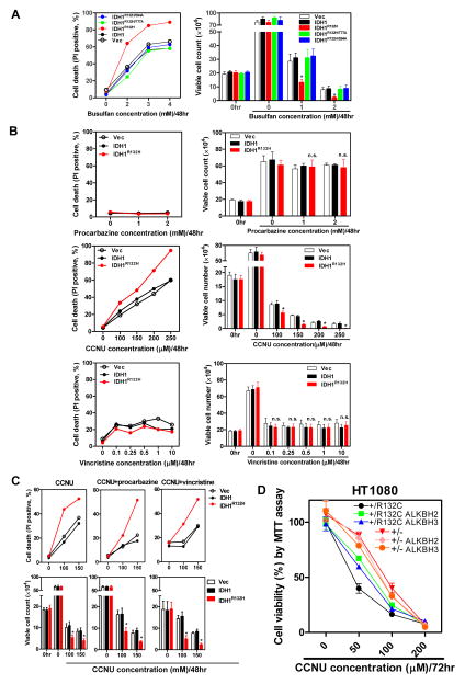
Figure 4. Expression of tumor-derived mutant IDH1 sensitizes cells to clinical alkylating agents.
(A, B) U87-MG cells stably expressing the indicated proteins were exposed to different concentrations of busulfan(A), procabazine, CCNU or vincristine (B) for 48 hrs. Cell viability was assessed by flow cytometry analysis (left), and cells were also stained with trypan blue for viable cell counting (right).
(C) U87-MG cells stably expressing the indicated proteins were treated with different concentrations of CCNU, along with 1 mM procarbazine or 500 nM vincristine for 48 hours. Cell viability was assessed by flow cytometry analysis (upper) and trypan blue exclusion (lower). Shown are average values of triplicated results with standard deviation (S.D.). *denotes the p < 0.05 for cells expressing mutant IDH1 versus wild-type IDH1; n.s.= not significant.
(D) Parental HT1080 (IDH1+/R132C) and TALEN-edited HT1080 (IDH1+/−) cells stably expressing ALKBH2 or ALKBH3 were exposed to different concentrations of CCNU for 72 hr. Cell viability was determined by MTT assay. *denotes the p < 0.05 for HT1080 (IDH1+/−) versus parental HT1080 (IDH1+/R132C) cells.

