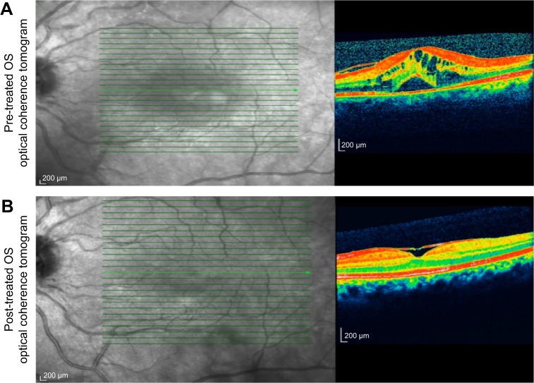
Figure 3.
Represents the spectral domain optical coherence tomography of the left eye at pre-treatment and post-treatment phases.
Notes: (A) Spectral domain optical coherence tomography of the patient with intermediate uveitis disclosed left cystoid macular edema with a foveal thickness of 693 μm associated with the retina pigment epithelial detachment and epiretinal membrane formation. (B) Spectral domain optical coherence tomography of the patient with intermediate uveitis disclosed regression of left cystoid macular edema and the retina pigment epithelial detachment with a foveal thickness of 362 μm associated with persistence of epiretinal membrane formation at the 3rd month of the MTX therapy.
Abbreviations: MTX, methotrexate; OS, left eye.

