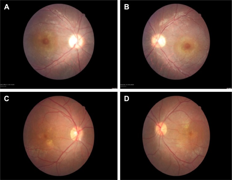
Figure 5.
Represents the color fundus photos of the right and left eyes.
Notes: Color fundus photos of right eyes (A and C) and left eyes (B and D) of two patients with focal chorioretinitis disclose yellowish lesions with faded centers at the level of the pigment epithelium in the macula and paramacular regions.

