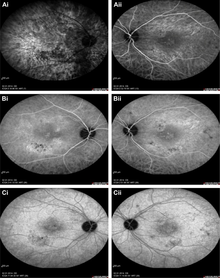
Figure 7.
Represents the indocyanine green angiography of the right and left eye.
Notes: Indocyanine green angiography early phase of right and left eyes with focal chorioretinitis disclosed areas of hypofluorescence (Ai and Aii), intermediate phase disclosed staining of choroid associated with areas of hypofluorescence (Bi and Bii), and late phase disclosed areas of hypo and hyperfluorescence associated with increased staining of choroid in the posterior pole (Ci and Cii).

