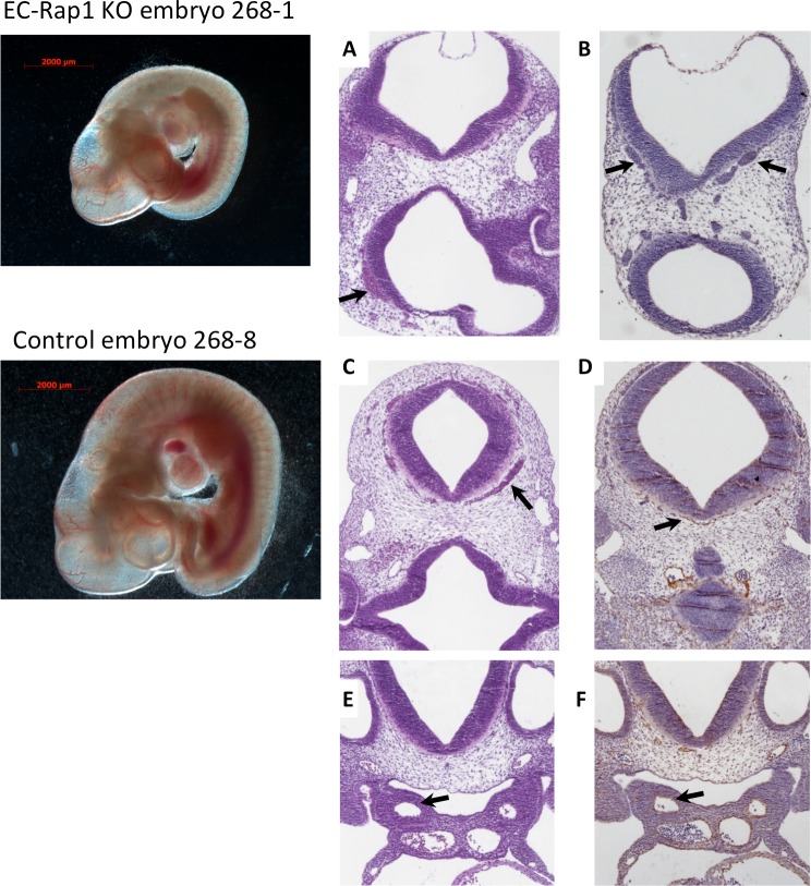
Fig 4. Normal branchial arches in EC-Rap1 KO embryos.
Left: whole mount images. Right: (A, B, top row): Dilated microvessels in E10.5 EC-Rap1 KO embryo heads near neural tube (arrows). C-F: Control (Tie2-Cre0; Rap1af/f, Rap1bf/f, E10.5) embryo sections. Blood-packed perineural vessels (C, D, arrows) are seen in fewer sections than in EC-Rap1KO embryos (B). (E, F, arrows) normal branchial arch arteries. Staining shown is H&E (A, C, E) and CD31 (B, D, F).

