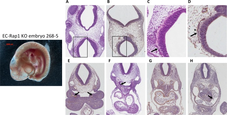
Fig 5. 50% of E10.5 EC-Rap1 KO embryos appear grossly normal.
Left: whole mount image. Right, A-D: normal perineural vasculature; C and D: enlargement of boxed areas in A and B; (C, arrow) blood-filled vessels; (D, arrow) ECs. E: 1st branchial arch artery, normal (arrow); F: larger 3rd branchial arch artery (arrow); G: aortae and cardinal veins with normal pericardial space at the level of atrium; (H, arrow) sinus venosus. Staining shown is H&E (A, C, F) and CD31 (B, D, E, G and H).

