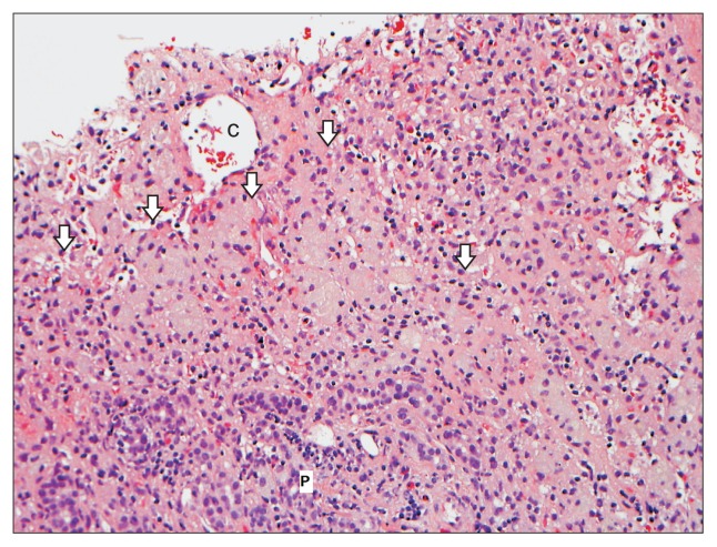
Fig. 3.
A liver biopsy from a 55-year-old female who developed a submassive hepatocyte necrosis and dropout 2 to 3 months after starting rosuvastatin. This photomicrograph shows large areas of hepatocyte dropout with multiple aggregates of reactive ceroid-laden macrophages (arrows). No viable hepatocytes are present in this picture (H&E stain, ×200).
C, central vein; P, portal tract.

