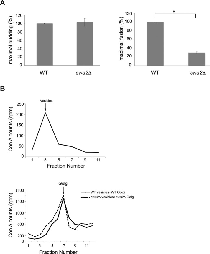
FIGURE 7:
The swa2Δ mutant is defective in COPII vesicle fusion but not budding or tethering in vitro. (A) Vesicle budding (left) and fusion (right) were measured in fractions prepared from wild type and the swa2Δ mutant as described in Materials and Methods. Error bars represent SEM, n = 3. *, p < 0.05, Student’s t test. (B) Top, fractionation of released transport vesicles: sucrose velocity gradient fractionation pattern of vesicles formed in vitro from wild-type permeabilized donor cells and cytosol. Bottom, vesicle tethering: the transport assay was performed with wild-type donor cells and a wild-type S1 fraction (black line) or swa2Δ mutant donor cells and a swa2Δ mutant S1 fraction (dotted line). At the end of the assay, the permeabilized donor cells were pelleted, and the supernatant was fractionated on a sucrose velocity gradient.

