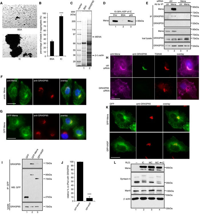
FIGURE 1:
Mena localizes to the cis-Golgi through interaction with GRASP65. (A) GRASP65- coupled Dynal M-500 beads were incubated with either buffer containing BSA or HeLa interphase cytosol (IC). After incubation, the beads were placed on glass slides, and random fields were photographed. Scale bar, 100 μm. (B) Quantification of A for the percentage of beads in aggregates. Results are expressed as mean ± SEM. Statistical significance was assessed by comparison to BSA-treated beads. The p value was determined by Student’s t test; ***p < 0.001. (C) Affinity purification of GRAS65-interacting proteins. Interphase cytosol fractionated by 15–30% ammonium sulfate precipitation was incubated with either BSA or His-GRASP65–coupled CNBr beads, and bound proteins were analyzed by SDS–PAGE and Coomassie blue staining. Arrows indicate the Mena and β/γ-actin bands that were excised and identified by mass spectrometry. (D) Beads coated with BSA or GRASP65 were incubated with 15–30% ammonium sulfate–precipitated proteins from interphase cytosol (I, input), washed, and analyzed by Western blot for Mena. (E) Immunoprecipitation (IP) by either nonspecific IgG (ctrl) or anti-Mena antibodies from lysate of cells transfected with control (ctrl) or Mena-targeting siRNA. (F) WT HeLa cells were immunostained with indicated antibodies to show the Golgi localization of endogenous Mena. (G) HeLa cells expressing GFP-Mena were immunostained for GRASP65. (H) Cells transfected with control or GRASP65 siRNA were immunostained for the indicated proteins. GRASP65 depletion abolished the Golgi localization of Mena. (I) Cells transfected with GFP, GFP-Mena, or GFP-VASP were lysed and immunoprecipitated by GFP antibodies. (J) Quantification of the amount of GFP-Mena or GFP-VASP that was coimmunoprecipitated with GRASP65, with the level of GFP-Mena normalized to 100% . ***p < 0.001. (K) Cells expressing GFP-Mena or GFP-VASP were immunostained for GRASP65. Mena but not VASP is concentrated on the Golgi. Bar, 20 μm (F–H, J). (L) Purified rat liver Golgi (RLG) membranes were incubated with interphase (IC) or mitotic (MC) cytosol or sequentially incubated with MC and then IC (MC → IC), reisolated, and blotted for indicated proteins.

