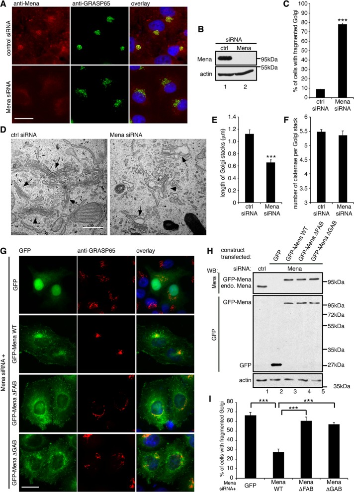
FIGURE 2:
Mena regulates Golgi integrity through actin polymerization. (A) HeLa cells treated with indicated siRNA were stained for Mena and GRASP65. (B) Immunoblot of HeLa cells from A. (C) Quantification of A for the percentage of cells with fragmented Golgi. (D) Representative EM images of the cells described in A. Arrows, Golgi stacks. Bar, 500 nm. (E) Quantification of D for the average cisternal length of Golgi stacks. (F) Quantification of D for the average number of cisternae per Golgi stack. (G) Mena-depleted HeLa cells were transfected with a cDNA for GFP or GFP-tagged, RNAi-resistant Mena WT or mutants and stained for GRASP65. Bar, 20 μm (A, G). (H) Cells were transfected first with nonspecific (lane 1) or Mena-specific siRNA (lanes 2–5) and then with plasmid of GFP (lane 2), GFP-tagged, RNAi-resistant Mena WT (lane 3), ΔFAB (lane 4), and ΔGAB (lane 5) mutants and analyzed by Western blot. endo. Mena, endogenous Mena. (I) Quantification of the percentage of GFP-positive cells with fragmented Golgi in G. ***p < 0.001.

