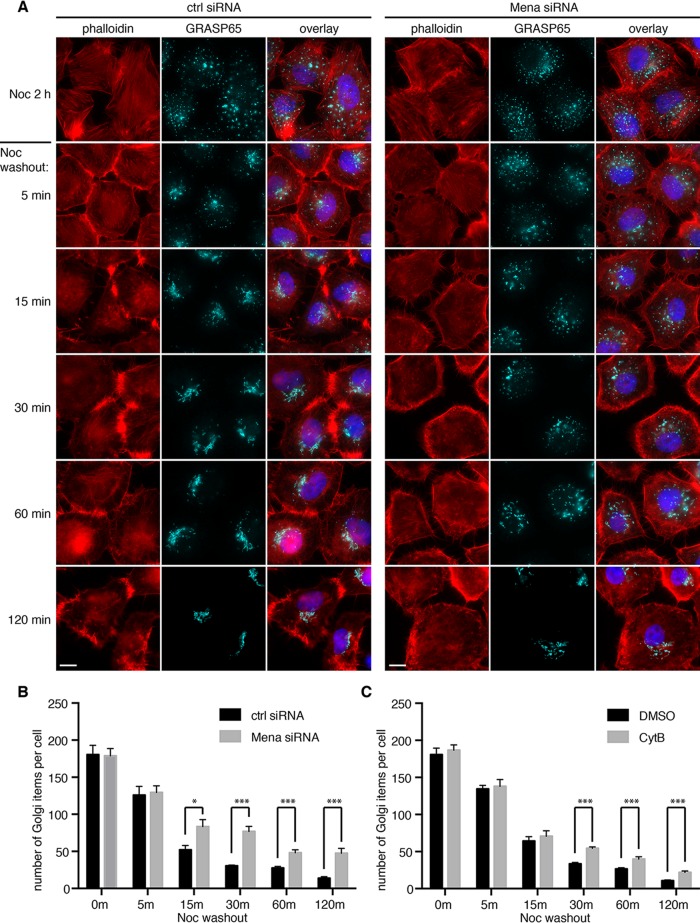
FIGURE 5:
Mena depletion impairs Golgi reassembly after nocodazole washout. (A) HeLa cells transfected with control or Mena siRNA were incubated with nocodazole for 2 h. After nocodazole removal for indicated times (minutes), cells were fixed and stained by phalloidin and GRASP65 antibodies. Bar, 10 μm. (B) Experiments in A were analyzed by confocal microscopy, and the number of Golgi particles in the indicated cells was quantified (mean ± SEM). *p < 0.05; ***p < 0.001. (C) Cells were incubated first with nocodazole for 2 h and then with the addition of DMSO or cytochalasin B for another 30 min. Cells were washed and further incubated in growth medium containing DMSO or cytochalasin B, but no nocodazole, for the indicated times. Cells were stained for GRASP65 and by phalloidin (images shown in Supplemental Figures S4 and S5) and analyzed by confocal microscopy and quantified as in B. Quantitation results.

