FIGURE 3:
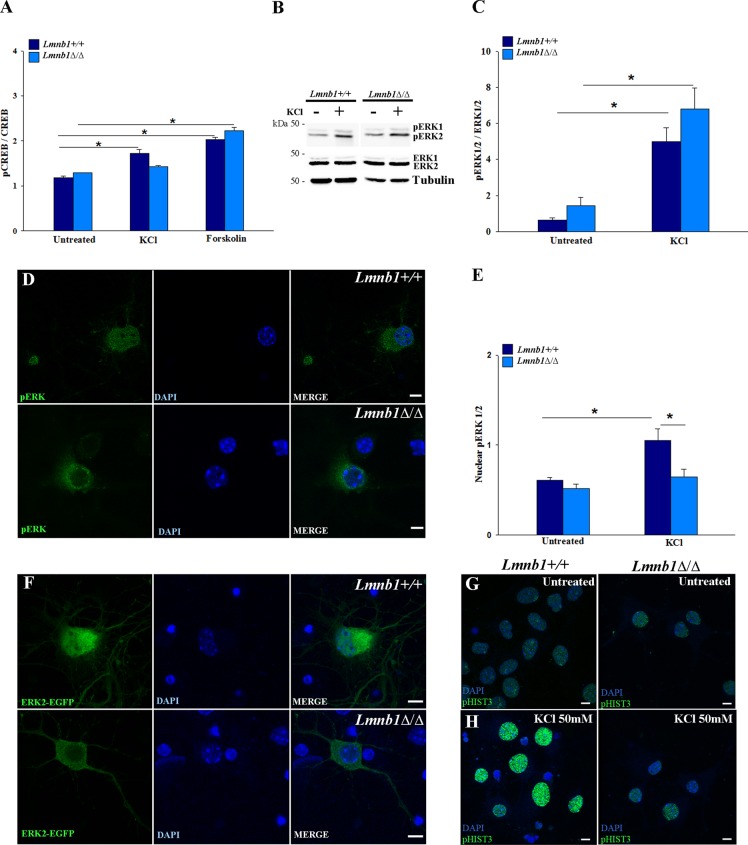
Lmnb1 deficiency impairs pERK nuclear import in mouse cortical neurons. Lmnb1+/+ and Lmnb1Δ/Δ neurons (7 DIV) were incubated with 50 mM KCl or 72 μM forskolin for 1 h. ERK, pERK, CREB, and pCREB were analyzed as described in Materials and Methods. (A) Quantitative analysis of pCREB immunoreactivity in the nucleus. Bars represent the average intensity ± SEM of nuclear pCREB immunofluorescence normalized to nuclear CREB immunofluorescence. At least 150 neurons/group were analyzed in two independent experiments. *p < 0.01, two-way ANOVA, followed by multiple comparison with the Holm–Sidak method. (B) Representative Western blots of ERK1/2, pERK1/2 and tubulin in neurons incubated with or without KCl. (C) Quantitative analysis of pERK levels. The data are normalized to total ERK. Bars represent the average ratio ± SEM from seven independent experiments. (D, E) Nuclear translocation of pERK in KCl-stimulated neurons. (D) Representative maximal projections of z-stack confocal images of pERK1/2 immunoreactivity (green). (E) Quantitative analysis of pERK immunoreactivity in the nucleus. Bars represent the average intensity ± SEM of nuclear pERK1/2 immunofluorescence normalized to cytosolic pERK1/2 immunofluorescence. At least 65 neurons/group were measured in three independent experiments. *p < 0.01, two-way ANOVA, followed by multiple comparison with the Holm–Sidak method. (F) pERK2-EGFP nuclear translocation. Representative maximal projections of z-stack confocal images of Lmnb1+/+ and Lmnb1Δ/Δ neurons transfected with pERK2-EGFP and incubated with KCl 50 mM for 1 h. (G, H) Representative maximal projections of confocal z-stack images of pHIST3 (green) immunoreactivity in 7DIV Lmnb1+/+ (left panels) and Lmnb1Δ/Δ (right panels) primary cortical neurons incubated in the absence (G) or presence (H) of 50 mM KCl for 1 h. In all fluorescence images, nuclei are counterstained with DAPI (blue). Scale bars, 5 μm.

