FIGURE 4:
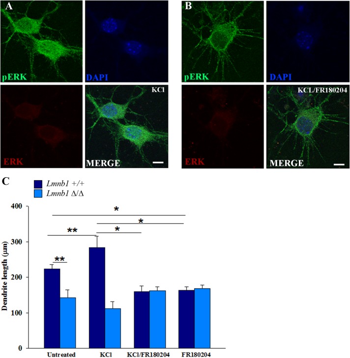
pERK-induced dendritic outgrowth is impaired in Lmnb1-deficient cortical neurons. pERK nuclear translocation and dendritic outgrowth in primary cortical neurons treated with or without KCl alone or in combination with FR180204. (A, B) Representative maximal projection of confocal z-stack images of pERK immunoreactivity (green) in Lmnb1+/+ primary cortical neurons (7 DIV) treated for 1 h with 50 mM KCl alone (A) or KCl and 36 μM FR180204 (B). Nuclei are counterstained with DAPI. Scale bars, 5 μm. (C) Quantitative analysis of total dendrite length of Lmnb1+/+ and Lmnb1Δ/Δ neurons incubated with or without 50 mM KCl or 36 μm FR180204 alone and in combination for 72 h. Bars represent mean total dendrite length ± SEM from four independent experiments. At least 45 neurons were analyzed for each experimental condition. *p < 0.05, **p < 0.01, two-way ANOVA, followed by multiple comparison with the Holm–Sidak method.

