FIGURE 6:
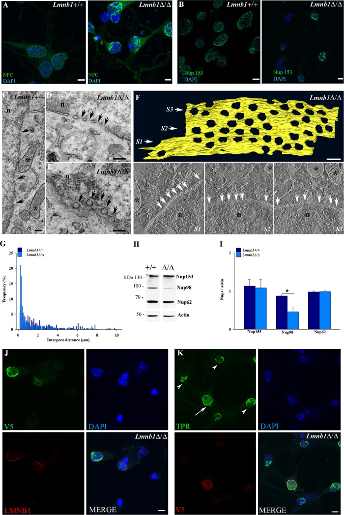
Lmnb1 deficiency alters the distribution of NPCs in mouse cortical neurons. NPCs were analyzed in 7 DIV Lmnb1+/+ and Lmnb1Δ/Δ primary cortical neurons as described in Materials and Methods. (A, B) Representative maximal projections of z-stack confocal images of Lmnb1+/+ and Lmnb1Δ/Δ neurons stained for NPCs (A; green) and Nup153 (B; green). (C–E) TEM images of Lmnb1+/+ (C) and Lmnb1Δ/Δ (D, E) neurons showing NPCs (arrows) in transversal (C, D) and paratangential (E) sections. Asterisks indicate mitochondria. n, nucleus. Scale bars, 200 nm. (F) Top, 3D models of a Lmnb1Δ/Δ nuclear membrane fragment representing the reconstruction of an 800-nm tomogram acquired in HAADF STEM from three semithin serial sections (Supplemental Movie S1). Scale bars, 200 nm. Bottom, single tomogram slices corresponding to sections S1–S3 in the three-dimensional model. White arrows point to NPCs; asterisks indicate mitochondria. n, nucleus. (G) Frequency distribution of interpore distances in nuclei of Lmnb1+/+ (n = 198) and Lmnb1Δ/Δ (n = 557) neurons. The frequency of interpore distances was determined using electron micrographs from >30 transversal sections. (H) Representative Western blot of NPCs and actin in lysates from Lmnb1+/+ and Lmnb1Δ/Δ cultured neurons. (I) Quantitative analysis of NPC expression. The data are normalized to actin. Bars represent the average ratio ± SEM. n = 4 per genotype. *p < 0.05, Student’s t test. (J, K) Lmnb1Δ/Δ neurons were infected with LV syn-tagged-V5-LMNB1 and analyzed by immunocytochemistry. (J) Representative maximal projection of confocal z-stack images of V5 (green) and LMNB1 (red) immunoreactivity in Lmnb1Δ/Δ neurons infected with LV syn-tagged-V5-LMNB1. (K) Representative maximal projection of confocal z-stack images of Tpr (green) and V5 (red) immunoreactivity in Lmnb1Δ/Δ neurons infected with LV syn-tagged-V5-LMNB1. In all fluorescence images, nuclei are counterstained with DAPI (blue). Scale bars, 5 μm.

