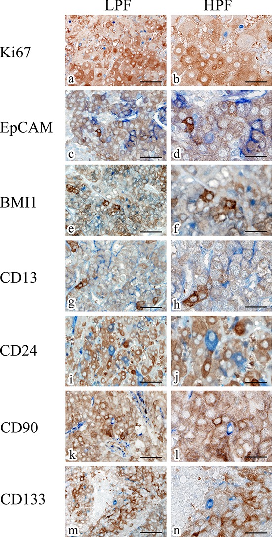
Figure 6. Representative double-staining immunohistochemistry for ALDH1A1 (brown) and Ki67 (blue), EpCAM (blue), BMI1 (blue), CD13 (blue), CD24 (blue), CD90 (blue) and CD133 (blue) in HCC specimens.

Scale bars indicate 100 μm (a, c, e, g, i, k, m) and 50 μm (b, d, f, h, j, l, n), respectively. Abbreviations: LPF, low-power field; HPF, high-power field.
