
Figure 3. Glioblastoma neovascularization and invasion depend on IRE1α catalytic activities.
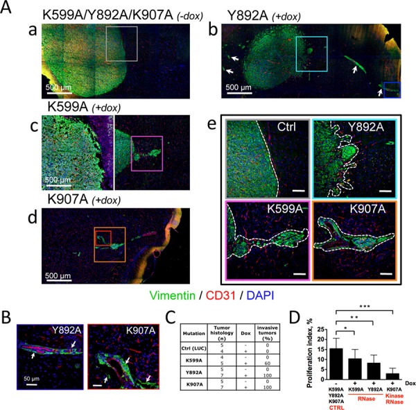
Glioblastoma cells bearing IRE1α mutants were xenografted into mouse brains and animals were fed with (+dox) or without (−dox) doxycycline. A. Invasive behavior of U87-K599A, U87-Y892A and U87-K907A-cell-derived tumors under doxycycline treatment. Aa-e. Coronal sections of mouse brains at day 28 (a, b, c) and day 47 (d) of tumor development were co-labeled using anti-vimentin (malignant cells, green labeling) and anti-CD31 (blood vessels, red labeling) antibodies. DAPI-labeled nuclei are in blue. Aa) Control tumors were massive and angiogenic. Ab) U87-Y892A cells developed into angiogenic and highly invasive gliomas. Perivascular glioma cell clusters (white arrows) were observed far away from the tumor core. Ac-d) Infiltrative phenotypes observed with U87-K599A cells and K907A cells. Ae) Higher magnification of boxed-areas in Aa-d. Dashed lines show borders between tumor tissues and normal brain tissues. (Scale bars: 100 μm.) B. U87-Y892A and U87-K907A glioma cells co-opted blood vessels. Higher magnifications of blue and red boxed-areas in Ab and Ad, respectively. C. Histological scoring of invasive tumors in each cohort. Any tumor responding to one of the three following criterias was scored as invasive: i) presence of a loosely delineated glioma rim, ii) existence of micro-satellites (n ≥ 10) in the periphery of the bulky core, and iii) emergence from the tumor core of extensions whose lengths were equal or superior to 500 μm. D. Measure of the proliferation index by using Ki-67 labeling. The percentage of dividing cells was obtained from the counting of at least 5, 000 nucleis for each tumor group (see also Figure S4A).