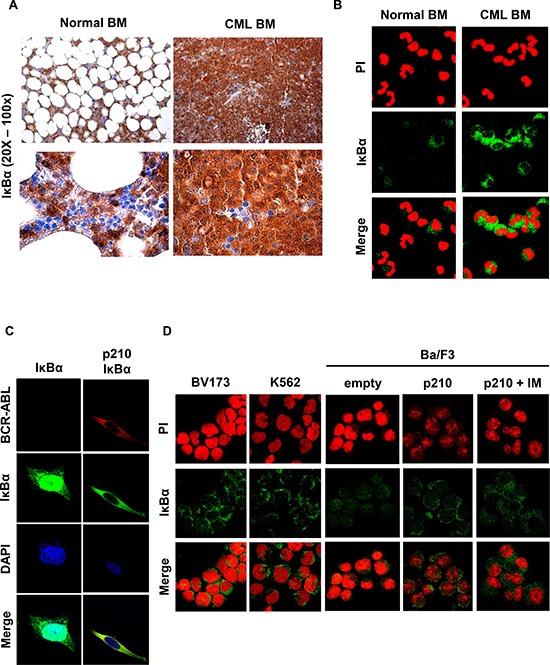
Figure 1. IκBα in BCR-ABL positive cells.

A. Representative immunohistochemistry using an anti-IκBα antibody performed on paraffin-embedded bone marrow from a representative CML or normal bone marrow. IHC was performed on 4 CML chronic phase samples with comparable results. B. Representative immunofluorescence on normal and CML primary bone marrow cells were performed to detect endogenous IκBα (green signal). Nuclei were stained with propidium iodide (red). Immunofluorescence was performed on 20 chronic phase CML samples and 3 normal bone marrow samples with comparable results. C. HeLa cells were transfected with BCR-ABL and IκBα plasmids. Immunofluorescence staining of IκBα (green) and BCR-ABL (red) was performed to detect IκBα localization. Nuclei were stained with DAPI. D. Immunofluorescence on the BV173 and K562 CML cell lines and on parental Ba/F3 and Ba/F3 p210 BCR-ABL cells was performed to detect IκBα (green signal). Nuclei were stained with propidium iodide (red). When indicated, cells were treated for 6 hours with 10 μM of imatinib.
