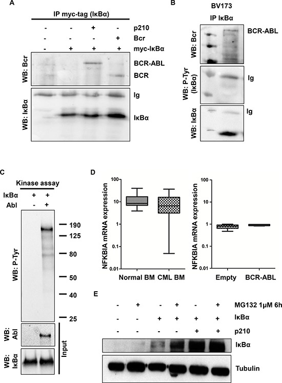
Figure 2. IκBα interactions and phosphorylation in BCR-ABL transfected cells.

A. HEK293T cells were transfected with the indicated plasmids and assessed by immunoprecipitation with myc-tag antibody (IκBα) and western blot. B. BV173 lysates were immunoprecipitated with IκBα antibody and western blot C. In vitro kinase assay with purified ABL and IκBα proteins was performed. P-Tyr: phospho-tyrosine. D. qRT-PCR analysis of NFKBIA (IκBα) performed on normal and CML bone marrow samples and on HEK293T BCR-ABL and empty vector transfected cells. E. BCR-ABL and IκBα-transfected HEK293T cells were incubated for 6 hours with 1 μM of MG132. Western blot analysis was performed to evaluate IκBα protein level. Tubulin was used as loading control.
