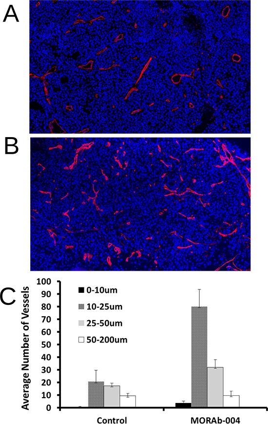
Figure 4. Immunofluorescent staining and digital analysis of tumor microvessels.

A. PBS treated (control) B16-F10 s.c. tumor section stained with Collagen IV (ColIV); B. MORAb-004 treated B16-F10 s.c. tumor section stained with ColIV; C. Comparison of digital counting of microvessels grouped by size. All images were captured at 20x using Panoramic Midi digital slide scanner.
