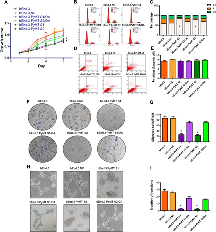
Figure 5. PyMT activates AKT and ERK leading to increased cell proliferation, migration and angiogenesis.

A. bEnd.3 cells displayed higher proliferation than bEnd.3 PyMT Si cells, and bEnd.3 PyMT Si cells regained rapid growth after treatment with OA. (n = 3/group, one-way ANOVA) *P < 0.05 B. Cell cycles analyzed via FACS. C. Obvious G1 cell arrest was observed in bEnd.3 PyMT Si cells compared with bEnd.3 cells, which could also be rescued by OA treatment. (n = 3/group, one-way ANOVA) *P < 0.05 D. Apoptosis was determined via AnnexinV and PI co-staining. E. No significant difference in the number of apoptotic cells was observed between bEnd.3 and bEnd.3 PyMT Si cells. F. Transwell assays demonstrated that PyMT silencing in bEnd.3 PyMT Si cells resulted in an approximately 70 percent decrease in migration. OA treatment could rescue this suppression effect. G. Quantitative analysis of cell migration. (n = 3/group, one-way ANOVA) *P < 0.05 H. In vitro angiogenesis tube formation assay results showed that bEnd.3 parental cells and bEnd.3 NC cells exhibited an ability to organize and form networks of cords on Matrigel after 48 h of culture, while PyMT-silenced bEnd.3 PyMT Si cells only formed islands of cells, with a few cells migrating out. Treatment with OA partially restored the angiogenic ability of bEnd.3 PyMT Si cells. I. Quantitative analysis of junctions number in angiogenesis tube formation assay. (n = 3/group, one-way ANOVA) *P < 0.05
