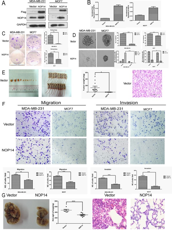
Figure 2. NOP14 overexpression inhibits tumorigenesis and metastasis of breast cancer cells.

A. Western blot analysis showing the exogenous expression of recombinant Flag-NOP14 protein, using primary antibodies against Flag and NOP14, respectively; and GAPDH expression was a loading control. B. qPCR showing mRNA transcription in stable cell lines with or without NOP14 constructs. C. Representative images of reduced foci formation in plate cultures. Quantitative analysis of foci numbers are shown in the right panel. Values reflect the mean ± 3SD of at least three independent experiments. D. Representative images of the decreased sphere forming ability of NOP14-expressing cells. The results are summarized as the mean ± 3SD of three independent experiments in the right panel. E. Tumor formation in nude mice demonstrates decreased in vivo tumorigenicity in NOP14-expressing cells (no tumor formed in the NOP14 overexpression group) compared with empty vector-transfected cells. F. Transwell assays and Matrigel invasion assays with representative image as upper panel and statistical results as the mean ± 3SD of three independent experiments in the lower panel. G. In vivo metastasis results with cell lines with empty vector or NOP14 construct. Panel for the left to the right are representative images of lung metastasis, wet weight and HE staining, respectively. (*P < 0.05, **P < 0.01, ***P < 0.001, independent Student's t test).
