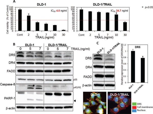
Figure 1. The mechanism of TRAIL-resistance was due to down-regulation of DR5 and malfunction of its recruitment.

A. TRAIL-sensitive DLD-1 and -resistant DLD-1 cells were treated with rTRAIL (2, 5, 7, 10, 30 ng/ml) for 24 h. The cell viability was estimated at 24 h after the treatment. Data were obtained from 3 independent experiments. The cell viability of the control (0; DMSO alone) is indicated as 100%. The growth inhibitory activity (IC50) of each compound is indicated in each panel. B. Western blot analysis was performed to determine the expression levels of DR5, DR4, FADD, caspase-8, and PARP-1 after the treatment with rTRAIL (5 and 7 ng/ml), with β-actin used as an internal control. C. Western blot analysis was performed to determine steady-state expression of DR5, DR4, and adaptor molecule FADD. β-actin was used as an internal control. Also shown are the steady-state expression levels of DR5 mRNA as relative ratios with respect to the GAPDH expression level. The expression level of mRNA was calculated by the ΔΔCt method. Means (S.D. indicated by error bars are shown. Lower photomicrographs: The photomicrograph shows the results of immunofluorescence staining for DR5 (anti-DR5) on the cell surface and in the cytosol of DLD-1 and DLD-1/TRAIL cells. Nuclei were counterstained in blue with Hoechst33342.
