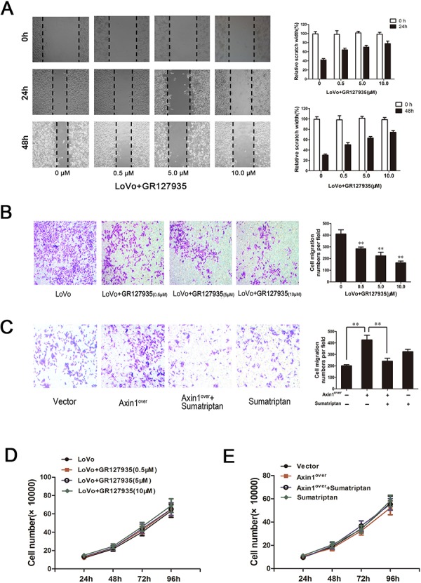
Figure 4. 5-HT1DR modulated invasion and migration in colorectal cancer cell.

A & D. Wound-healing assay. Images were taken 0, 24 and 48 hours after wound formation. Data are presented as mean ± SD of triplicate experiments.*P < 0.05, **P < 0.01 ##P < 0.01 in Fig. 4D. B & C. Cell invasion assay result using Matrigel-coated Transwell. (left, representative pictures of invasion chambers; right, average counts from five random microscopic fields). Data are presented as mean ± SD of triplicate experiments. *P < 0.05 vs. Vector group in Fig. 4A; *P < 0.05, **P < 0.01 in Fig. 4B. D.The cell proliferation of GR127935-treated LoVo cells in different concentration at 24 h, 48 h, 72 h, 96 h as described in Materials. E. Cell proliferation was assayed at 24, 48, 72 and 96 hours after LoVo cells were transfected with Axin1 lentivirus (Axin1over) or treated with sumatriptan or treated with both together.
