
Figure 1. LIF, LIFR and gp130 immunohistochemical expression in CCA and peritumoral areas of human liver samples.
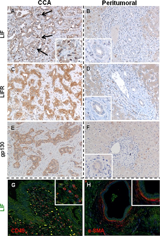
In CCA bile ducts, the extensiveness of LIF A. expression was heterogeneously distributed amongst samples, whilst the staining of LIFR C. and gp130 E. was more homogeneous. In contrast, LIF B., LIFR D. and gp130 F. immunoreactivity was significantly less in bile ducts of matched peritumoral tissues. By immunohistochemistry and dual immunofluorescence we demonstrate that LIF (green) was also extensively expressed by CD45+ inflammatory cells (red, G.) and α-SMA+ cells (CAF, red, H.) that juxtaposed neoplastic biliary structures (A, black arrows and inset, G, and H) (Original magnification: A-H, 200 ×; insets, 400 ×).