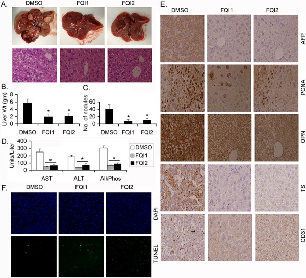
Figure 1. LSF inhibitors abrogate endogenous HCC in Alb/c-myc mice.

Protocols for induction of HCC and treatment of animals are described in Materials and Methods. A. Upper panel, representative photographs of livers of DMSO-, FQI1- and FQI2-treated mice at the end of the experiment. Lower panel, representative H & E stained liver sections of the indicated group at the end of the experiment. Magnification: 400X. B. Liver weight of the mice in the indicated treatment groups. C. Number of liver nodules in the indicated treatment groups. D. Serum levels of aspartate aminotransferase (AST), alanine aminotransferase (ALT) and alkaline phosphatase (Alk Phos) in the indicated treatment groups. For B-D, n = 10 in each group. The data represent mean ± SEM. *:p < 0.01. E. Immunohistochemical analysis of the indicated proteins in the liver sections of the indicated groups. Arrows indicate microvessels. Magnification: 400X. F. TUNEL staining in the liver sections of the indicated groups.
