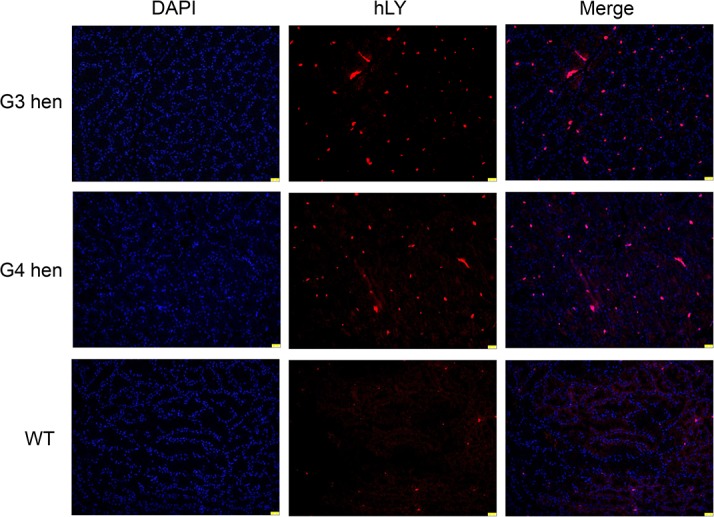
Fig 2. Immunofluorescence detection of hLY expression in oviduct sections.

Sections of the magnum portions of the oviducts from a G3, a G4, and a wild type White Leghorn hen were immunolabeled with the anti-hLY antibody (red). The nuclei were stained with DAPI (blue). Scale bar: 25 μm.
