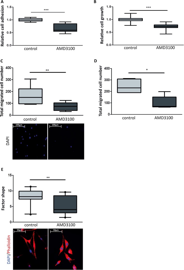
Figure 5. Effect of an allosteric CXCR4 inhibitor on breast cancer cell growth and migration.
A. Box plots illustrating the relative cell adhesion of MDA-MB-231GFP_luc cells. Treatment of breast cancer cells with AMD3100 (10 μM) decreases relative cell adhesion to irradiated Beas-2B cell monolayer. n = 12; ***, P < 0.001(Unpaired t-test with Welch's correction). B. Box plots illustrating the relative cell growth of MDA-MB-231GFP_luc cells. Treatment of breast cancer cells with AMD3100 (10 μM) decreases relative cell growth in presence of CMLE-IR. (Unpaired t-test with Welch's correction). n = 15; ***, P < 0.001 (Unpaired t-test). In C. and D., quantification by bioluminescent imaging after 4 days incubation. Data is represented as relative fold change compared with the corresponding control value. C. Box plots illustrating impact of AMD3100 (10 μM) on total migrated cell number of MDA-MB-231GFP_luc cells stimulated by CMLE-IR. Nuclei were stained blue with DAPI (lower panel). n = 9; **, P = 0.009 (Unpaired t-test with Welch's correction). D. Box plots illustrating total transendothelial migrated cell number of MDA-MB-231GFP_luc cells stimulated by CMLE-IR in presence of AMD3100 (10 μM) or control. n = 6; *, P = 0.017 (Mann-Whitney U). E. Box plots illustrating the extent of CMLE-IR -induced cell spreading of MDA-MB-231GFP_luc cells treated with AMD3100 (10 μM) versus control, as quantified by factor shape (upper panel). Fluorescence microscopy images of cells double stained with phalloidin for actin filaments (red) and DAPI counterstaining for nuclei (blue) (lower panel). n = 20; **, P = 0.004 (Mann-Whitney U).

