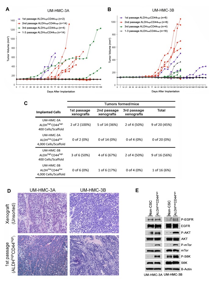
Figure 3. Tumorigenic potential of low passage mucoepidermoid carcinoma cells sorted for ALDH/CD44.
A., B. Graphs depicting tumor volume of A. UM-HMC-3A or B. UM-HMC-3B xenograft cells FACS-sorted for ALDH/CD44. Scaffolds were seeded with either 400 ALDHhighCD44high or 4,000 ALDHlowCD44low cells and transplanted into the subcutaneous space of SCID mice. Existing tumors were retrieved, re-sorted and 400 ALDHhighCD44high or 4,000 ALDHlowCD44low cells seeded into new scaffolds, and serially passaged in vivo. C. Table depicting the number of tumors grown in the ALDHhighCD44high versus ALDHlowCD44low populations for each passage performed. D. H&E staining of tumors generated with FACS-sorted ALDHhighCD44high and ALDHlowCD44low cells. Images were taken at 100X. E. UM-HMC-3A and UM-HMC-3B cells were sorted for ALDHhighCD44high or combined ALDHhighCD44low, ALDHlowCD44high, and ALDHlowCD44low (non-CSC population). NP-40 lysis buffer was used to prepare whole cell lysates that were resolved using PAGE. Membranes were probed using antibodies a 1:1000 dilution against human mTor, p-mTor, Akt, p-Akt, S6K, p-S6K, p-EGFR; 1:2000 dilution of EGFR, and beta-actin.

