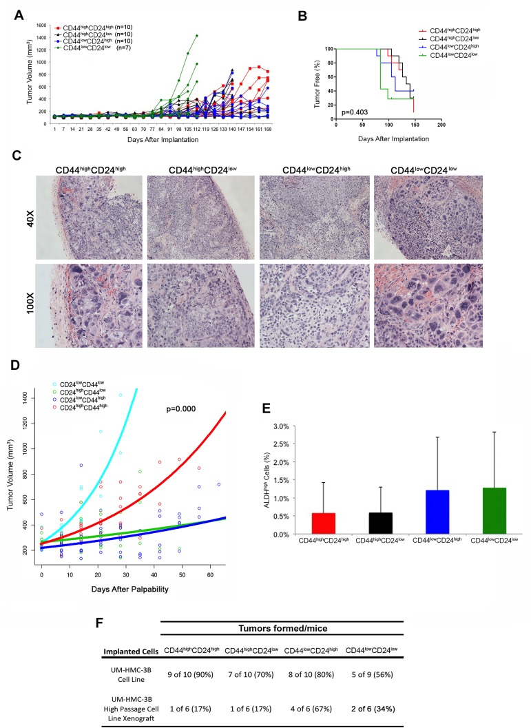
Figure 6. Tumorigenic potential of mucoepidermoid carcinoma cells sorted for CD44/CD24.
A. In vivo transplantation of 5,000 UM-HMC-3B (passage 103) FACS-sorted cells (CD44highCD24high, CD44highCD24low, CD44lowCD24high, or CD44lowCD24low) with 900,000 endothelial (HDMEC) cells seeded on biodegradable scaffolds and transplanted into the subcutaneous space of SCID mice. B. Kaplan-Meyer analysis of time to palpability of tumors generated with cell sorted for CD44/CD24. Tumors were considered palpable once they reached 200 mm3. C. H&E staining of tumors generated by the transplantation of UM-HMC-3B cells sorted for CD44/CD24. Images were taken at 40X and 100X. D. Regression analysis of growth after palpability (200 mm3) of tumors generated with cells FACS-sorted for CD44 and CD24. E. Graph depicting the percentage of ALDHhigh cells in tumors generated with cells FACS-sorted for CD44/CD24. F. Table depicting the number of tumors formed in each CD44/CD24 sorted sup-population in both the UM-HMC-3B cell line and the UM-HMC-3B low passage cell line xenograft model.

