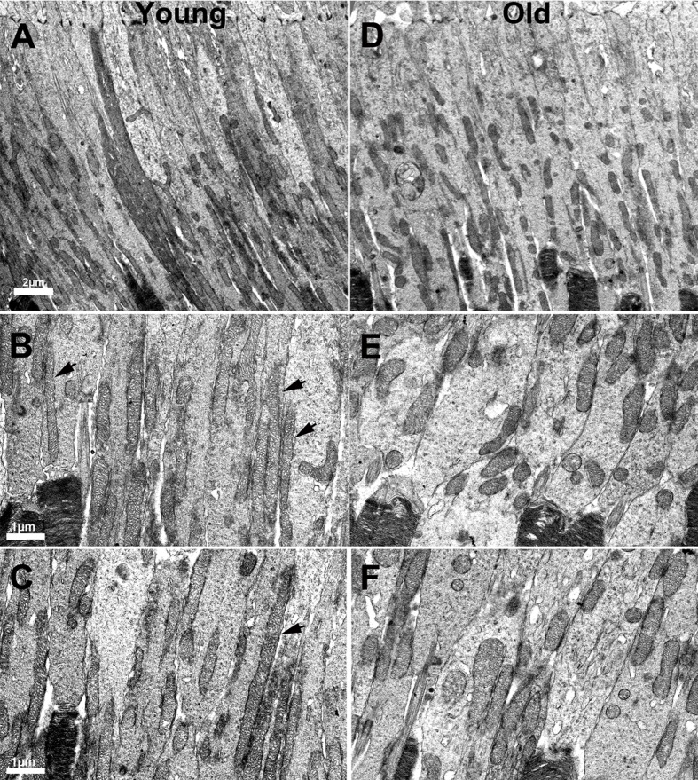
Figure 5. Changes in the morphology of mitochondria in ageing.
A. Low power electron micrograph from young retinae showing elongated and tubular like mitochondria present in photoreceptor inner segments. B.–C. Electron micrograph from young retinae at higher magnification showing thin tubular and elongated mitochondria present in the inner segment along the membrane (black arrows). D. Electron micrograph of low magnification from an old mouse showing mitochondria that are relatively fragmented, being shorter in photoreceptor inner segments. E.–F. Electron micrograph from old mice showing fragmented mitochondria that are shorter and fragmented compared to those found in young animals.

