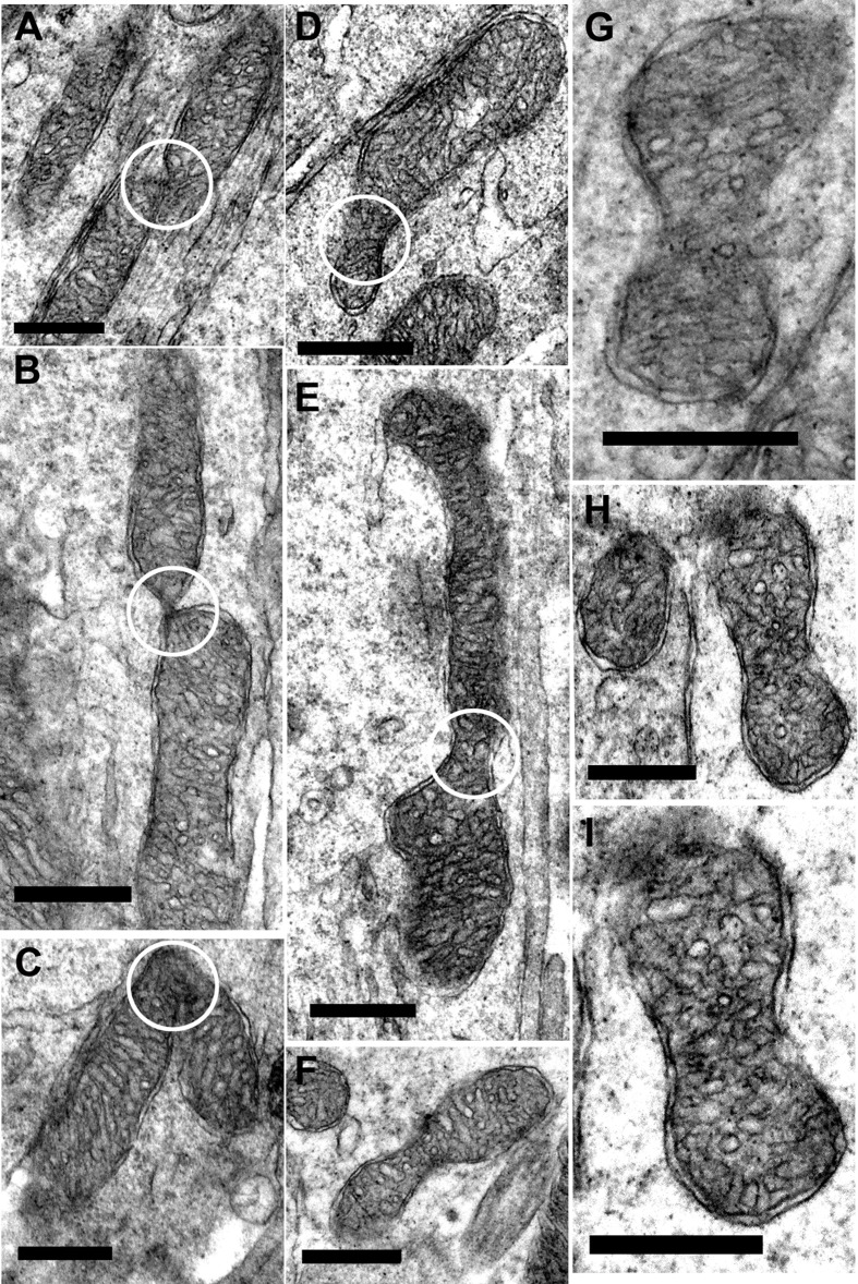
Figure 8. Fission and fusion in inner segment of old mice.
A.–C. Representative electron micrographs of mitochondria undergoing fission/fusion in the inner segment of old mice (white circle) where the mitochondrion is either dividing or fusing. D.–I. Electron micrographs of suspected mitochondrial fusion/dividing in the old mice. (White circle indicate the ‘pinching off’ of the mitochondria for either fission or fusion). Scale bar = 0.5 μm.

