
Figure 3. NIR laser can penetrate the mouse abdominal wall and cause an SPR effect of pSGNs to kill cancer cells in vivo.
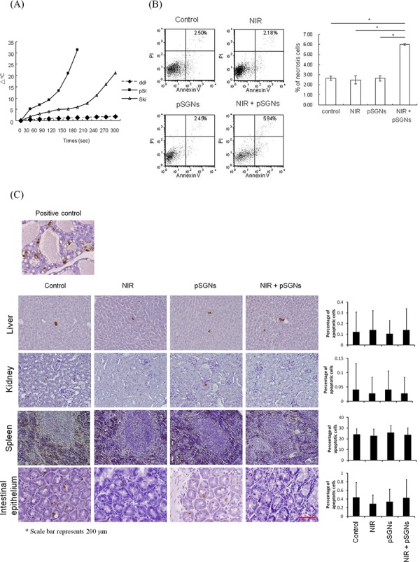
A. The abdominal skin of NOD/SCID mice was dissected and placed on a glass slide covering the 96-well plate. The abdominal skin was irradiated using a 2000-mW NIR laser, and the temperature of 100 μL of pSGNs (OD800 = 6) in the 96-well plate under the abdomen wall was monitored using a thermocouple. The wells that contained distilled water or pSGNs and were irradiated directly with the NIR laser without a mouse abdominal skin covering were used as control. The pSGNs under the mouse abdominal skin that were irradiated with the NIR laser generated heat and increased the solution temperature. These results imply that the NIR laser can penetrate the NOD/SCID mouse abdomen and cause pSGNs to generate heat through the SPR effect. B. 1 × 10⁶ ID8 cells were intraperitoneally injected into NOD/SCID mice. After 3 days, the mice were divided into four groups of five mice and intraperitoneally injected with 2 mL of pSGNs (OD800 = 1.5) or 10% trehalose and irradiated with an 808-nm NIR laser (3.2 W/cm2) in five different areas on the abdomen for a total of 5 min. One day after the final NIR irradiation, the mice implanted with ID8 cells were sacrificed, and i.p. lavage was performed to collect cancer cells. The lavaged cells were then stained with annexin V and propidium iodide. GFP-positive cancer cells were gated, and annexin V-PI double-positive necrosis cells were analyzed using flow cytometry. No significant difference in the percentage of necrosis in i.p. cancer cells was observed among control, NIR-laser irradiated, and i.p. pSGNs-only groups. However, the percentage of necrosis in cancer cells was significantly increased in the groups that received i.p. photothermal treatment mediated by pSGNs. C. The normal tissues from the mice of each group were dissected and fixed in 10% formalin. The damage of normal tissues in the intraperitoneal cavity was evaluated using a TUNEL assay, and the percentage of damaged cells was determined using ImageJ software. Normal female rodent mammary gland tissue, 3 to 5 days after weaning of rat pups, was used as positive control. The size of the scale bar was 200 μm. This result demonstrated that photothermal therapy did not cause noticeable damage in normal tissues in vivo. The error bar represents the standard error. (P < 0.01*)