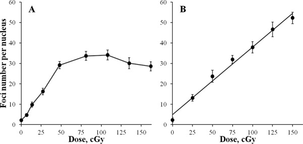
Figure 1. Formation of γH2AX foci in diploid normal human fibroblasts during continuous exposure to X-ray radiation at a dose-rate of 4.5 mGy/min A.

or 30 min after acute X-ray irradiation B. γH2AX foci were quantified using immunofluorescence microscopy. Two hundred cells per data point were analyzed per experiment. Means calculated from three independent experiments ± standard errors are shown.
