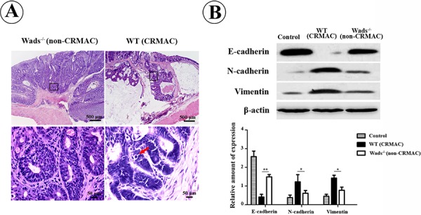
Figure 5. CRMAC in WT mice showed more invasiveness than non-CRMAC in Wads−/− mice did at week 37.

A. Tumor cells and mucinous extended beyond the basement membrane of mucosa and invaded into submucosa in WT mice, in which tumors developed more atypical glands and displayed more pathologic mitoses (red arrow). Views in the frames of upper panel are zoomed in in lower panel. B. EMT was strikingly elevated in CRMAC compared with non-CRMAC indicated by decreased E-cadherin and increased N-cadherin and vimentin. (*P < 0.05,**P < 0.01).
