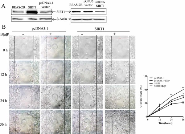
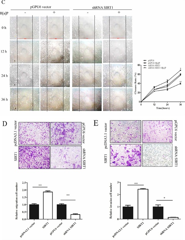
Figure 3. SIRT1 promoted BEAS-2B cells migration and invasion upon B[a]P exposure.


A. BEAS-2B cells were stably transfected with pcDNA3.1 vector/pcDNA3.1-SIRT1 or pGPU6 vector/shRNA-SIRT1. The success of transfection was confirmed by Western blotting. The whole-cell extracts were probed with anti-SIRT1 antibody. B. Cell migration behaviors in the stable pcDNA3.1 vector/pcDNA3.1-SIRT1 transfected BEAS-2B cells were evaluated by wound-healing assay. The pictures were taken at different time. C. Wound healing assay of pGPU6 vector/shRNA-SIRT1 transfected cells. Quantification(closure rate%) were obtained from three independent experiments and expressed as mean ± S.D. (n = 3). *p < 0.05 and **p < 0.01. D–E. Cell migration and invasion ability were analyzed by transwell assay. Results were expressed as mean ± S.D. (n = 3). The numbers of cells were obtained from three independent experiments. **p < 0.01.
