
Figure 6. IWR-1 suppression of the EMT in the ex vivo model of colorectal cancer.
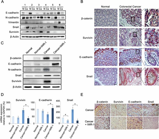
A. Western blot analysis showing that colorectal cancer (CRC) specimens exhibited the increased survivin expression and EMT-like expressional changes (decreased E-cadherin and increased N-cadherin and Snail expressions) compared to the matched controls. B. Immunohistochemical analysis of CRC tissues showing the increase of β-catenin (purple color), survivin (purple-to-red color), and Snail (red color) and decrease of E-cadherin (disappearing of red color) (Magnification, ×100, scale bar 50 μM). Generally, positive expressions of β-catenin, survivin, E-cadherin, and Snail were stained in purple-to-red colors. C. Western blot analysis showing IWR-1 effects on EMT markers in CRC tissues. IWR-1 significantly decreased the protein expressions of β-catenin and survivin, and inhibited EMT in CRC tissues. D. RT-qPCR showing IWR-1 effects on the mRNA expressions of the EMT markers in CRC tissues. IWR-1 significantly decreased the mRNA expressions of β-catenin and survivin, and inhibited EMT in CRC tissues. E. Immunohistochemical stains demonstrating that IWR-1 increased E-cadherin and decreased survivin and Snail expressions in CRC tissues.