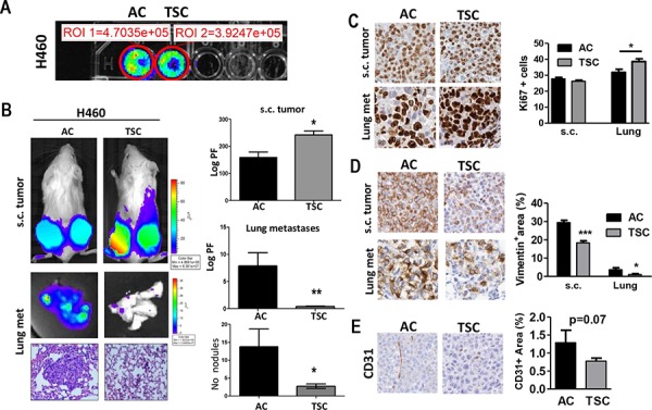
Figure 2. TSC cells display higher tumorogenic potential and decreased metastatic activity.

A. In vitro BLI of adherent cultured (AC) cells and disaggregated tumor sphere cultured (TSC) cells for H460 cell line (20 000 cells per well). B. Left panels: Representative images of BLI of total body (upper panel) and excised lungs upon necropsy, and representative H&E stained lung sections for both groups (bottom panel). Two groups of mice were subcutaneously (s.c.) injected with H460 cells (TSC and AC). Right panels: Quantification of BLI (Photon flux: Log PF) showing increased tumorigenic potential of TSC cells after subcutaneous injection into mice (upper panel), while AC cells led to an increased lung tumor burden (middle panel) and the number of lung nodules (lower panel) in lung histological sections. Error bars are mean ± SEM. (p < 0.05). C. Representative IHC for Ki67+ in resected s.c. tumors and lung metastases in TSC versus AC-injected mice. Right panel: Quantification of proliferating cells in both s.c. tumors and lung metastases. D. Representative vimentin staining in s.c. tumors (left panel) and quantification (right panel) for both s.c. tumors and lung metastases, showing reduced positive area in TSC-derived tumors. E. IHC staining and quantification for CD31+ in s.c. tumors showing a trend towards a decrease in angiogenesis but it did not reach statistical significance. *p, 0.05; **p < 0.01; ***, p < 0.001), Error bars are mean ± SEM.
